
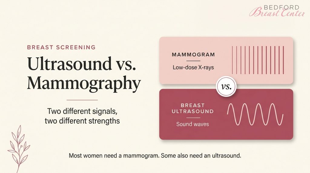
A normal mammogram doesn’t always tell the whole story. Dense tissue shows up white on an X-ray, and so does a tumor, so some cancers can hide in plain sight.
That’s why the ultrasound versus mammography question matters for you.
A mammogram remains the standard screening test. Still, a breast ultrasound can fill gaps, especially with dense breasts or implants. The right choice in a breast ultrasound vs mammogram comparison depends on your age, tissue type, and history.
At Bedford Breast Center, we weigh those factors, plus any implants you have, then recommend the screening that fits you.
Key Takeaways
- Ultrasound versus mammography is about purpose. Mammograms screen the whole breast; ultrasound checks one area.
- Mammograms detect tiny calcium deposits, a possible early cancer sign that ultrasound often misses.
- Breast ultrasound vs. mammogram: ultrasound separates fluid-filled cysts from solid lumps and uses no radiation.
- Dense breasts and implants can obscure findings, so you may need both tests.
- The USPSTF recommends mammograms every two years for average-risk women ages 40 to 74.
When Should You Start Breast Cancer Screening?
If you are at average risk, plan to start mammograms at 40. Stanford Medicine researchers estimate that mammography and better treatment together cut US breast cancer deaths by 58% between 1975 and 2019. Finding cancer early also gives you more treatment options.
In April 2024, the US Preventive Services Task Force finalized a recommendation for mammograms every two years for ages 40 to 74. The American Cancer Society makes yearly screening optional from 40 to 44, then recommends it from 45 to 54.
At Bedford Breast Center, we recommend yearly screening starting at age 40.
Age also shapes the ultrasound versus mammography decision. If you are under 30 and find a lump, ultrasound usually comes first because younger breast tissue is dense. Breast self-exams help you notice changes, but they cannot replace imaging.
What Is a Breast Ultrasound?
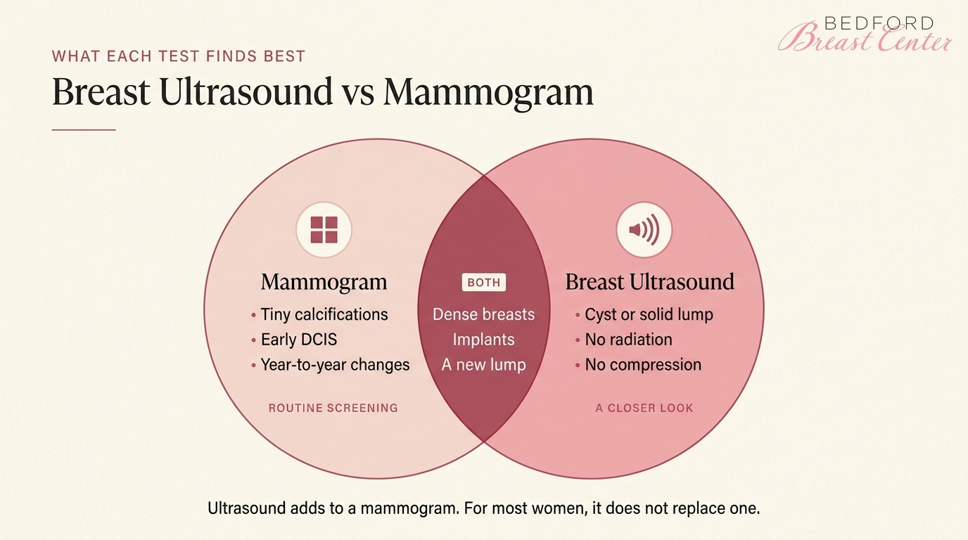
A breast ultrasound uses high-frequency sound waves to image the tissue inside your breast. Unlike a mammogram, it involves no radiation and no compression, which is the main difference between breast ultrasound and mammography.
Your technologist spreads clear gel on your skin, then moves a handheld probe over the area being checked.
For most women, ultrasound is a follow-up or add-on test rather than a routine screen. If a mammogram shows something unusual, or you feel a lump, your doctor will usually order one next.
A handheld exam takes up to about 30 minutes. Automated breast ultrasound (ABUS) scans your whole breast with a larger probe, and the Breast Cancer Research Foundation notes the FDA approved it as a secondary screening method if you have dense breasts.
Ultrasound Advantages
- No radiation, so it is safe during pregnancy and easy to repeat
- Shows whether a lump is a fluid-filled cyst or a solid mass
- Produces real-time images, so a radiologist can guide a needle biopsy
- Can reveal some cancers hidden in dense tissue
- Comfortable, with no breast compression
Ultrasound Limitations
Ultrasound gives you a detailed view of one area, but it leaves gaps that a mammogram fills.
It can miss microcalcifications, tiny calcium deposits that sometimes signal early cancer. MD Anderson also notes that ultrasound is less effective at finding stage 0 breast cancer (DCIS), which is why it is not recommended as a replacement for a mammogram.
- Images only the area scanned, which makes year-to-year comparison harder
- Produces more false positives, which can lead to extra imaging or biopsies
- Depends on the skill of the person performing the scan
- Can be less accurate in very large breasts
What Is a Mammogram?
A mammogram is a low-dose X-ray of your breast. Each breast is gently compressed between two plates to image the tissue well, and a screening exam typically takes two views of each breast.
Mammography is also the best tool for spotting microcalcifications, small calcium deposits that can sometimes signal early cancer. Because the images cover your whole breast, your radiologist can compare them with earlier exams.
That whole-breast view is why mammography leads ultrasound versus mammography for routine screening.
At Bedford Breast Center, we use 3D mammography, which builds a three-dimensional picture so small tumors hidden on 2D images are easier to see. You are also less likely to be called back. If you are, a diagnostic mammogram adds extra views of the area that needs a closer look, and a radiologist reviews them while you are there.
Mammogram Advantages
- Images the entire breast in a few minutes.
- Best test for calcium deposits and early cancers, including DCIS.
- Shows asymmetry and distortion in breast tissue.
- Creates a baseline your radiologist can compare against every year.
- The only imaging test proven to save lives through screening, according to MD Anderson.
Mammogram Limitations
- Uses a small dose of radiation.
- Less sensitive in dense breast tissue, where tumors and dense tissue both look white.
- Brief compression can feel uncomfortable.
- False alarms can lead to callbacks and extra imaging.
Breast Ultrasound vs Mammogram: The Differences at a Glance
In a breast ultrasound vs mammogram comparison, each test answers a different question. This table sets them side by side:
|
Mammogram |
Breast ultrasound |
|
|
How it works |
Low-dose X-rays |
High-frequency sound waves |
|
Radiation |
Yes, a low dose |
None |
|
Compression |
Yes, for a few seconds |
No |
|
What it images |
The whole breast |
A targeted area (or the whole breast with ABUS) |
|
Best at finding |
Calcifications, early cancers, changes over time |
Cysts vs solid lumps, masses hidden in dense tissue |
|
Typical role |
Routine screening |
Follow-up, dense breasts, lumps, under 30, pregnancy |
The two tests work best as a pair, which is why ultrasound versus mammography is rarely an either-or choice. For example, a British Columbia study of more than 5,200 women with dense breasts found that adding ultrasound to mammography detected 6.1 additional cancers per 1,000 screens.
Your age and breast density also change the picture. A 2009 study of 546 women with breast symptoms found ultrasound more sensitive than mammography under age 45, while mammography performed better over 60. Density mattered too: mammography lost sensitivity as tissue became denser, while ultrasound stayed more sensitive.
Ultrasound vs Mammogram for Dense Breasts and Breast Implants
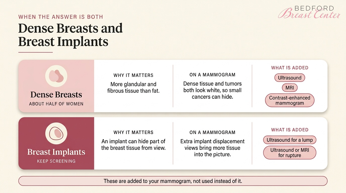
Dense tissue and breast implants are the two situations where ultrasound versus mammography most often ends with "both."
Dense Breasts
Dense breasts have more glandular and fibrous tissue than fat, and about half of women have them.
On a mammogram, dense tissue and tumors both appear white, which makes small cancers harder to spot. Dense tissue also carries a slightly higher cancer risk.
Since September 10, 2024, the FDA has required every mammogram report to include your breast density, and your patient summary now says if your tissue is dense. If it is, the notice adds that other imaging tests may help find cancers, so ask your doctor about supplemental options:
- Breast ultrasound, the add-on we recommend for dense tissue
- MRI, considered case by case based on your overall risk
- Contrast-enhanced mammography, which uses contrast dye to highlight areas of increased blood flow
Keep your annual mammogram either way, because these tests add to it rather than replace it.
Ultrasound vs Mammogram With Breast Implants
Ultrasound vs mammogram for breast implants is rarely an either-or choice, because the two tests answer different questions.
Most women with implants should still get regular screening mammograms, so tell the technologist about your implants before the exam starts. They can then take implant displacement views, which push the implant back and pull your breast tissue forward to image more of it.
Ultrasound helps in two situations:
- You feel a lump near an implant.
- Your doctor wants to check the implant itself.
For silicone implants, the FDA says ultrasound is an acceptable alternative to MRI for finding a silent rupture when you have no symptoms. Ask your surgeon how often to schedule it. Either way, you keep your mammograms and add ultrasound when a specific question comes up.
Mammogram or Ultrasound: Which Do You Need?
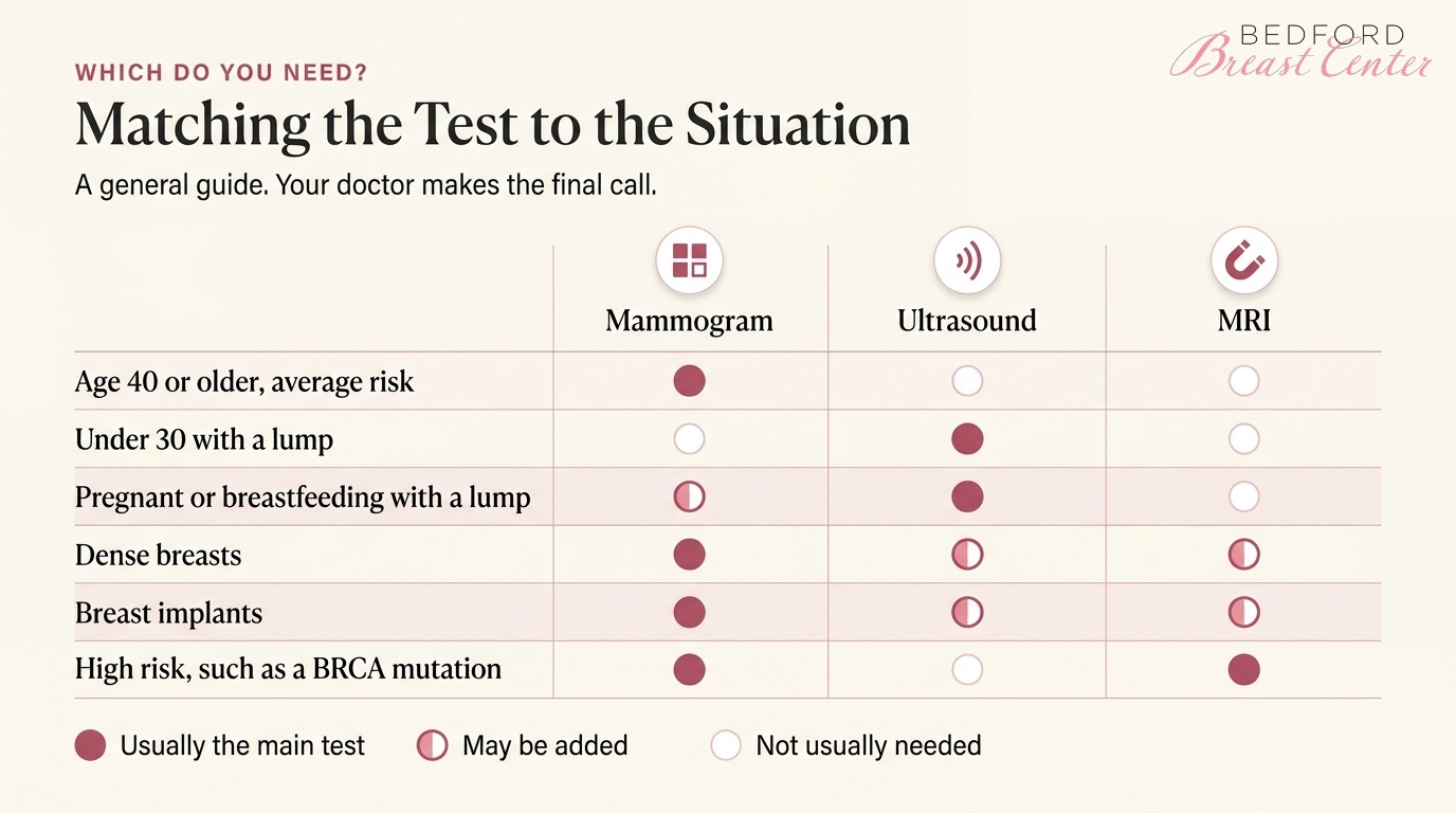
The ultrasound versus mammography answer depends on your situation:
- You are 40 or older at average risk: a mammogram, yearly or every other year.
- You are under 30 with a lump: ultrasound, usually before any mammogram.
- You are pregnant or breastfeeding with a lump: ultrasound first, then a shielded mammogram if needed.
- You have dense breasts: a mammogram, plus a talk about supplemental ultrasound, MRI or contrast-enhanced mammography.
- You have implants: a mammogram with implant displacement views, plus ultrasound or MRI if needed.
- You are at high risk: with a BRCA mutation or a lifetime risk of about 20% to 25% or more, the American Cancer Society recommends a yearly MRI and mammogram from age 30, and genetic testing can confirm a BRCA mutation.
Your doctor makes the final call, and your plan changes if your symptoms do.
Contact Our Beverly Hills Breast Medical Center Today
Bedford Breast Center offers 3D mammography and breast ultrasound in Beverly Hills. If your mammogram shows an abnormal mass, we can handle the follow-up ultrasound.
Unsure about ultrasound versus mammography for your own breast screening? Our team can walk you through your options. Call us at (310) 278-8590 Monday to Friday, 9 a.m. to 5 p.m., or contact us online to book your screening.
FAQs
Is a Mammogram an Ultrasound?
No. A mammogram uses low-dose X-rays to image your whole breast, while a breast ultrasound uses sound waves to examine a specific area. In ultrasound versus mammography, the mammogram is the standard screening test. Ultrasound usually follows up on a finding or adds a closer look when you have dense breasts.
Can Ultrasound Replace a Mammogram?
For most women, no. In a mammogram vs ultrasound comparison, mammography finds microcalcifications that ultrasound can miss, and it is the only imaging proven to save lives through screening. Ultrasound works best as an add-on for dense breasts, lumps or implants, and your doctor decides when you need both.
Ultrasound vs Mammogram by Age: Which Is Better Under 40?
Your age shapes the ultrasound vs mammogram choice. If you are under 30 and feel a lump, ultrasound usually comes first because younger breast tissue is dense. At average risk, you start screening mammograms at 40. With a high genetic risk, your doctor may add MRI from age 30.
Does a Breast Ultrasound Use Radiation?
No. A breast ultrasound uses high-frequency sound waves, so you have no radiation exposure and no compression. That is why it is also the first test for a lump during pregnancy. A mammogram uses a low-dose X-ray, which is the main practical difference between a breast ultrasound and a mammogram.
Ultrasound vs Mammogram With Breast Implants: Which Do You Need?
You still need screening mammograms with implants, taken with extra implant displacement views. For ultrasound vs mammogram breast implants, ultrasound is an add-on for a lump or an implant check. The FDA says ultrasound or MRI can also screen silicone implants for silent rupture. Ask your surgeon how often.
