Breast ultrasound is often done at our Beverly Hills center to check abnormal mammogram results. This diagnostic test uses sound waves to determine whether a breast lump is filled with fluid (a cyst) or is solid.
Early detection through comprehensive breast screening is key when treating breast cancer. According to the American Cancer Society, the 5-year survival rate is approximately 99% when breast cancer is localized and detected early.
A thorough breast cancer screening is the best tool for early detection. When most women consider breast cancer detection, they automatically think of mammograms. A mammogram is absolutely necessary to evaluate abnormal breast tissue, but a breast ultrasound can be just as important in the diagnostic process.
“My Obgyn recommended Bedford Breast Center for my breast ultrasound and I am EXTREMELY PLEASED with the level of attentiveness and care. They answered all of my questions during my very first breast ultrasound and made me feel comfortable, valued and safe to ask all questions in regards to my breasts. Thank you so much. I highly recommend.”
Kristen C. – Google
What Is a Breast Ultrasound?
A breast ultrasound is a safe, painless imaging test that uses sound waves to determine whether a breast lump is a solid mass or filled with fluid (a cyst). Breast ultrasounds do not use radiation. Instead, sound waves produce an image of the breast’s internal structures. While a breast ultrasound doesn’t replace the need for a mammogram, this form of breast screening can check for abnormal results.
A breast ultrasound is currently the only noninvasive test to determine if an abnormal mass is a cyst. The only other way to diagnose a cyst is through a breast biopsy, which requires removing the fluid through a needle.
Let’s Take A Closer Look At 3 Types Of Breast Imaging Tests
This Is How We Use An Ultrasound Machine For Breast Cancer Screening
What Is Sonocine Ultrasound And Is It Worth It?
Screening vs. Diagnostic Imaging | Here’s What You Need To Know
3 Imaging Test We Use to Screen for Breast Cancer
What Does Breast Cancer Look Like on an Ultrasound?
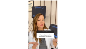
Why Would You Need an Ultrasound of the Breast?
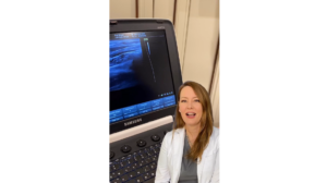
Ultrasound Technology 101
Supplemental Screening Options
Let’s Take A Closer Look At 3 Types Of Breast Imaging Tests
This Is How We Use An Ultrasound Machine For Breast Cancer Screening
What Is Sonocine Ultrasound And Is It Worth It?
Screening vs. Diagnostic Imaging | Here’s What You Need To Know
3 Imaging Test We Use to Screen for Breast Cancer
What Does Breast Cancer Look Like on an Ultrasound?
Why Would You Need an Ultrasound of the Breast?
Ultrasound Technology 101
Supplemental Screening Options
Why Are Breast Ultrasounds Used?
Breast ultrasounds are often performed to evaluate issues detected during a routine physical exam or mammogram. A breast ultrasound is the appropriate next step if your mammogram detects an abnormal mass or your physician feels a lump during a manual breast exam.
While most masses are benign, early detection of breast cancer dramatically improves overall outcomes. In women with a breast cancer diagnosis, a breast ultrasound is used to examine the lymph nodes under the arms, indicating whether the cancer has spread.
Breast ultrasound is especially valuable for younger women, who naturally have dense breast tissue and abundant milk glands. Dense glandular tissue can make it difficult to distinguish normal structures from possible tumors on a traditional mammogram. In these situations, ultrasound provides clearer visualization.
What Are the Limitations of a Breast Ultrasound?
A breast ultrasound is an effective tool for examining breast tissue, but it isn’t a comprehensive test. That’s why a mammogram is also necessary for a detailed analysis. Breast ultrasounds cannot:
Show microcalcifications: A microcalcification is a buildup of calcium around a tumor that can be telltale signs of cancerous tumors.
Capture images of the entire breast: Breast ultrasounds are taken through handheld transducers, so capturing an image of the entire breast in one snapshot is impossible.
Evaluate deep breast tissue: Breast ultrasounds primarily evaluate tissue closer to the surface of the breast. For deeper tissue inspection, a mammogram is recommended.
Unparalleled Expertise With a Compassionate Touch
Dr. Richardson and Dr. Memsic are two of the top breast surgeons in Los Angeles, using innovative techniques to create excellent clinical and aesthetic outcomes.
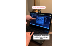
During an ultrasound, you will lie on an exam table and raise your arms over your head. Once you’re comfortable, the sonographer will apply a special gel to your breasts and slowly move the handheld transducer over each one.
The sound waves bounce off the breast tissue while the transducer picks them up. The reflected sound waves are transformed into images, allowing your doctor to view the tissue inside your breasts.
During the test, your sonographer can attach a Doppler probe device to the transducer to hear the sound waves. A Doppler probe helps your provider listen to the blood flow within the blood vessels of your breast tissue. If the flow is faint or silent, it may indicate a blockage.
Does a Breast Ultrasound Hurt?
Breast ultrasounds should never feel painful or uncomfortable. However, you may feel slight discomfort if the transducer passes over a sensitive or tender area of your breast. Because ultrasound technology doesn’t use radiation, they are safe during pregnancy.
“The ultrasound tech who I have seen for years is always incredibly kind and professional. I have never felt like I was being rushed through. They take their time with exams and are always available to answer your questions.”
K.H. – Google
Is There Any Downtime After My Breast Ultrasound?
Most breast ultrasounds take about 30 minutes to complete. In most instances, you can resume your normal activities immediately after the exam.
What Is an Ultrasound-Guided Biopsy?
If your breast ultrasound or mammogram detects a suspicious mass, your doctor can perform an ultrasound-guided biopsy. During this in-office procedure, the ultrasound helps direct the doctor to the exact area of concern. The ultrasound displays an image of your breast tissue so the doctor can efficiently reach the mass.
During a biopsy, the doctor inserts a tiny needle into the mass. Next, they remove a small sample of cells, which a pathologist later analyzes. This type of biopsy is less invasive than a surgical biopsy, with little to no scarring.
Trust Our Breast Surgery Experts
Our world-class facility in Beverly Hills serves patients from Los Angeles from Los Angeles, Santa Monica, Glendale, Malibu, and throughout Southern California. To learn more, call us at
(310) 278-8590 or contact us using the online form to schedule an appointment.